Blog
Q&A: Prasanthi Yerra on CMOS image sensors for surgical views
A Sensors Converge 2026 preview…
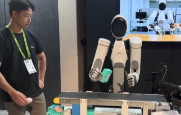
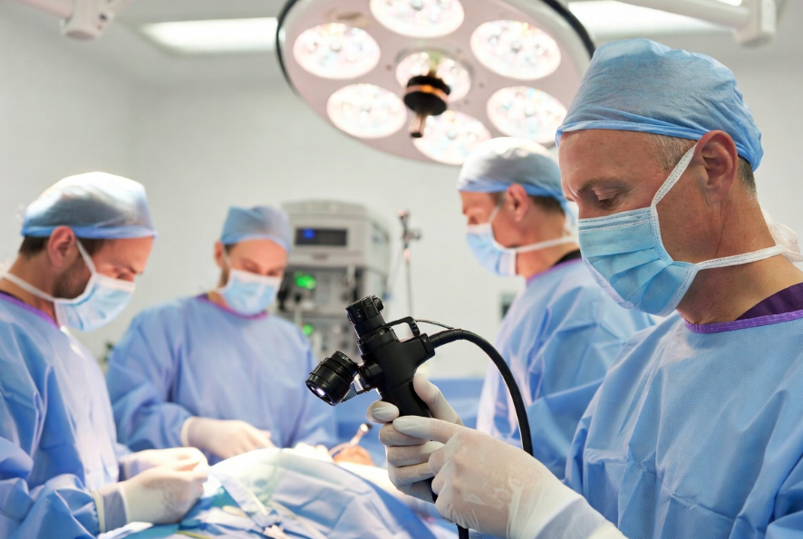
CMOS image sensors are seemingly everywhere–from cameras used to shoot striking images of the far side of the Moon to endoscopes used in delicate surgeries.
Fierce Sensors caught up with an expert in safely isolating data links needed for real-time high-resolution image sensors used in invasive medical devices.
Prasanthi Yerra has seven years experience in electronic applications development at Analog Devices as Systems Application Engineer. Her background includes a Master’s Degree in Electronic Engineering and she has since worked on IoT projects and Laser Beam Scanning technology. Her current focus is systems application of precision signal chain and isolation products.
She speaks on the topic: “Cost-Efficient Isolated Data Link for Real-Time High-Res Image Images in Invasive Medical Devices” on Thursday, May 7, 9:30 a.m. at Sensors Converge 2026 at Santa Clara Convention Center.
Fierce: Presanthi, what’s the essence of your talk addressing cost-efficient isolated data links in medical imaging?
Yerra: Surgical visualization has evolved dramatically. Imaging systems have transitioned from rod lens, CCD, and fiber-based technologies to advanced CMOS image sensors positioned at the tips of endoscopes and surgical tools. These sensors now deliver multi-stream 4K video critical for complex surgeries.
But here’s the challenge: when a camera is patient-connected, you must electrically isolate multi-gigabit video streams to meet IEC 60601-1 safety standards. Currently there’s no off-the-shelf solution for isolating the MIPI CSI-2 interface these sensors use. Our session presents two cost-efficient architectures: FPGA-based ones for maximum flexibility, and Bridge IC-based for streamlined production that combines Analog Devices’ GigaSpeed iCoupler isolation with GMSL connectivity. Ultimately, we are enabling customers to adopt newer system architectures that reduce overall costs while significantly increasing imaging performance.
Fierce: Can you share market insights on the history of CMOS sensors in surgical tools and how much the market has grown?
Yerra: The transformation over the past two decades has been substantial. We’ve moved from analog video and fiber-based imaging to CMOS-on-tip architectures that place high-resolution sensors directly at the distal end of surgical instruments. The market reflects this shift. The medical-grade CMOS sector is on pace to double by 2035, reaching up to $5.2 billion. While baseline growth sits at a steady 7 to 9% annually, the real engine is surgical imaging. Surging at over 10% a year, CMOS is actively displacing legacy CCD technology and now commands roughly 64% of the medical camera market.
This dominance comes down to three factors: lower power consumption, higher integration, and reduced manufacturing costs. CMOS is the enabling technology behind high-growth device categories like disposable endoscopes and robotic-assisted visualizations. We’re seeing rapid adoption of single-use endoscopic instruments in bronchoscopy and urology, driven by infection control requirements. This trend is reshaping design priorities toward simplified connectivity and reduced bill-of-materials cost.
The applications span gastroenterology, urology, laparoscopy, arthroscopy, and robotic-assisted procedures. Each has unique form factors and sterilization requirements, but they share a common need: reliable, high-bandwidth, safe camera interfaces.
Fierce: Why is electrical isolation so critical in surgical imaging, and what specific standards must medical device designers meet?
Yerra: When an endoscope or surgical camera contacts a patient, it’s classified as an “applied part” under IEC 60601-1. This requires two means of patient protection (2 MOPP) from hazardous voltages—a non-negotiable safety requirement. Isolation protects patients from electrical hazards while eliminating ground loops and confining noise in sensitive imaging electronics.
Fierce: What are the key technical challenges engineers face when designing isolated camera interfaces for surgical tools?
Yerra: Designing isolated camera interfaces for surgical tools involves a tightly coupled set of constraints. Bandwidths are scaling rapidly — a single 4K60 camera generates upwards of 12 Gbps of raw video, and multi-camera systems push well beyond that. At the same time, engineers must contend with significant signal loss over thin medical-grade cables, strict form-factor and thermal limits in the handpiece, and sterilization compatibility across multiple reprocessing cycles. Adding to this, MIPI CSI-2’s D-PHY — which switches between low-power and high-speed modes — is fundamentally incompatible with standard isolators. Addressing all these simultaneously requires a true system-level architecture rather than simple component selection.
This is where isolation technology becomes critical. Traditional fiber-based solutions add cost, complexity, and bulk. Our iCoupler technology portfolio takes a different approach: delivering 2 MOPP (Means of Patient Protection) at 50 Vrms per IEC 60601-1 with 10 Gbps total bandwidth, enabling gigabit-class isolation without fiber optics. It’s a purpose-built solution for the constraints surgical imaging demands.
Fierce: What will attendees walk away learning from your Sensors Converge session?
Yerra: Attendees will leave with a clear understanding of the system-level challenges behind MIPI CSI-2 isolation and actionable knowledge to overcome them. By exploring critical design constraints and GMSL signal chain integration, engineers will gain a direct path to implementation using our drop-in hardware platforms, providing the system-level solutions needed to define next-generation imaging architectures.
Prasanthi Yerra speaks at 9:30 a.m. PDT, May 7, at Sensors Converge 2026. Registration for Sensors Converge, May 5-7, is available online.